Portal Venous System Color Doppler Ultrasound
Evaluation of blood flow in the liver's portal vein, hepatic artery and veins for clots and narrowing.
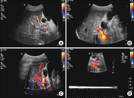
What is portal venous system color Doppler ultrasonography?
In a portal vein color Doppler ultrasonography examination, blood flow in the portal vein and hepatic artery branches feeding the liver, the hepatic veins leaving the liver, and the inferior vena cava (the main vein) is evaluated. Possible clot formations and narrowings in these vessels are investigated.
While the portal vein carries blood rich in digested nutrients coming from the spleen, pancreas, stomach and intestines, the hepatic artery brings oxygen-rich blood coming through vessels originating from the main artery (aorta) leaving the heart. The venous drainage of the liver occurs through the hepatic veins into the inferior vena cava.
Although vascular diseases of the liver are rare, their incidence has been increasing in recent years. In particular, portal vein diseases are important for human life because they can lead to acute liver failure, chronic liver disease and non-cirrhotic portal hypertension, and can even cause death.
Is preparation required for portal vein color Doppler ultrasound?
As with abdominal and hepatobiliary ultrasound, 6–8 hours of fasting is required before the examination to reduce the amount of gas in the abdomen, improve image quality and prevent some changes in blood flow.
Eating causes an increase in portal system pressure and enlargement of vessel diameter, which may change the results. Medications normally used can be taken with a little water. The patient does not need to have a full bladder.
How is portal vein color Doppler ultrasound performed?
In a portal venous system color Doppler examination, the patient lies on their back with the upper abdomen exposed. A harmless, transparent gel that facilitates the transmission of high-frequency sound waves is applied to the upper right abdomen, and imaging is performed with the ultrasound probe. The internal structure of the liver is examined in general with ultrasound.
The portal vein, hepatic artery and hepatic veins are examined for clots, vessel diameters are measured, and the speed and direction of the flows within the vessels are evaluated with color Doppler ultrasound imaging.
The blood flow in the vessel is displayed in red or blue on the ultrasound screen according to its direction. During the examination, the patient is asked to hold their breath from time to time. Holding the breath correctly and for a sufficient time makes it easier to measure the flow within the vessel effectively.
How long does portal vein color Doppler ultrasound take?
Portal vein color Doppler ultrasound takes approximately 20–30 minutes, and after the examination the patient can return to their daily activities. Color Doppler ultrasound examination has no known harm to the human body.
Is portal vein color Doppler ultrasound a painful examination?
Liver color Doppler ultrasound is a painless examination.
Does portal vein color Doppler ultrasound have any side effect or harm?
Liver color Doppler ultrasound has no known side effect or harm.